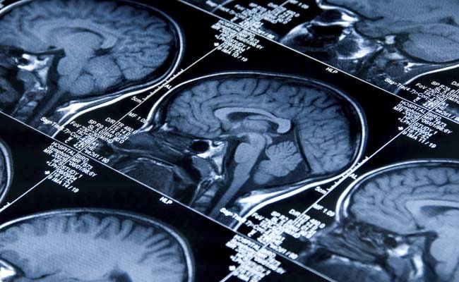
Photo for representational purpose.
Researchers have discovered that tracking changes in five brain areas linked to movement and balance with a simple non-invasive imaging technique could help evaluate experimental treatment to slow or stop the progress of Parkinson's.
The researchers used functional magnetic resonance imaging (fMRI) to reveal areas where Parkinson's disease and related conditions cause progressive decline in brain activity.
While current treatments focus on controlling symptoms, biomarkers provide a quantifiable way to measure how medications address not just symptoms, but the neurological changes behind them.
Previous studies have used imaging techniques that require the injection of a drug that crosses the blood-brain barrier.
"Our technique does not rely upon the injection of a drug. Not only is it non-invasive, it's much less expensive," said the study's senior author David Vaillancourt, Professor at University of Florida.
The researchers used functional MRI to evaluate five areas of the brain that are key to movement and balance.
A year after the baseline study, 46 Parkinson's patients in the study showed declining function in two areas -- the primary motor cortex and putamen. Some patients showed declines in all five areas.
The brain activity of the 34 healthy control patients did not change.
The finding, published in the journal Neurology, builds on a 2015 University of Florida study that was the first to document progressive deterioration from Parkinson's via MRI, showing an increase in unconstrained fluid in an area of the brain called the substania nigra.
(This story has not been edited by NDTV staff and is auto-generated from a syndicated feed.)
Track Latest News Live on NDTV.com and get news updates from India and around the world

